Your Cart is Empty
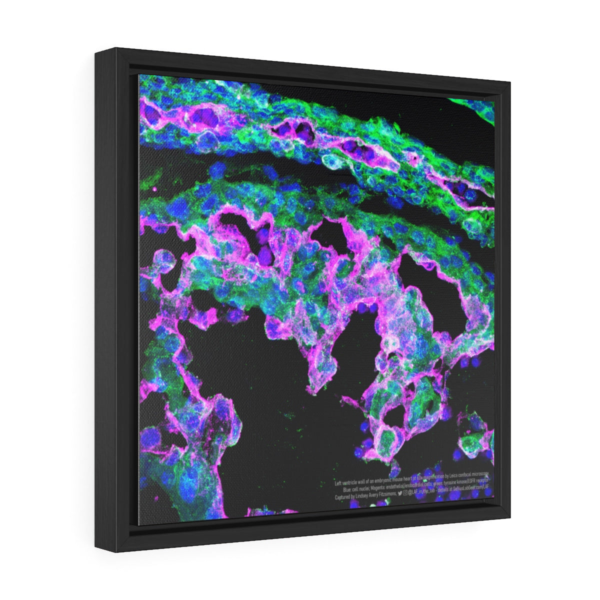
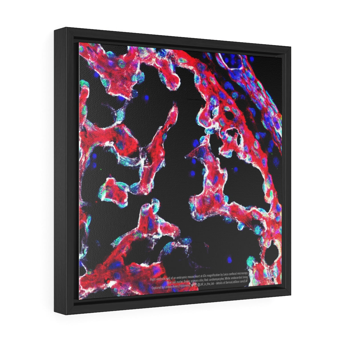
Left Ventricle Wall - Heart Microscopy by Lindsey Fitzsimons
$68.50 - $79.50
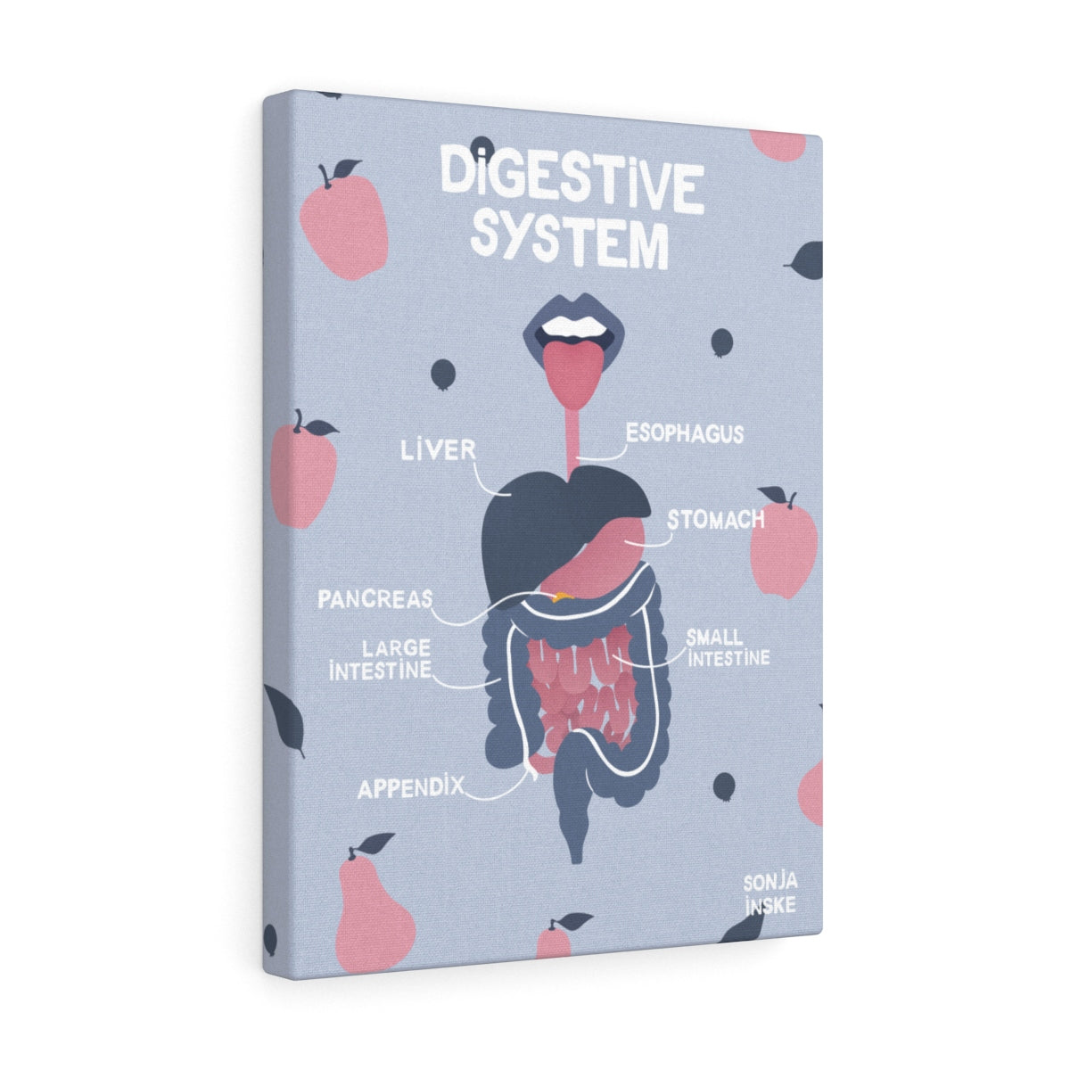
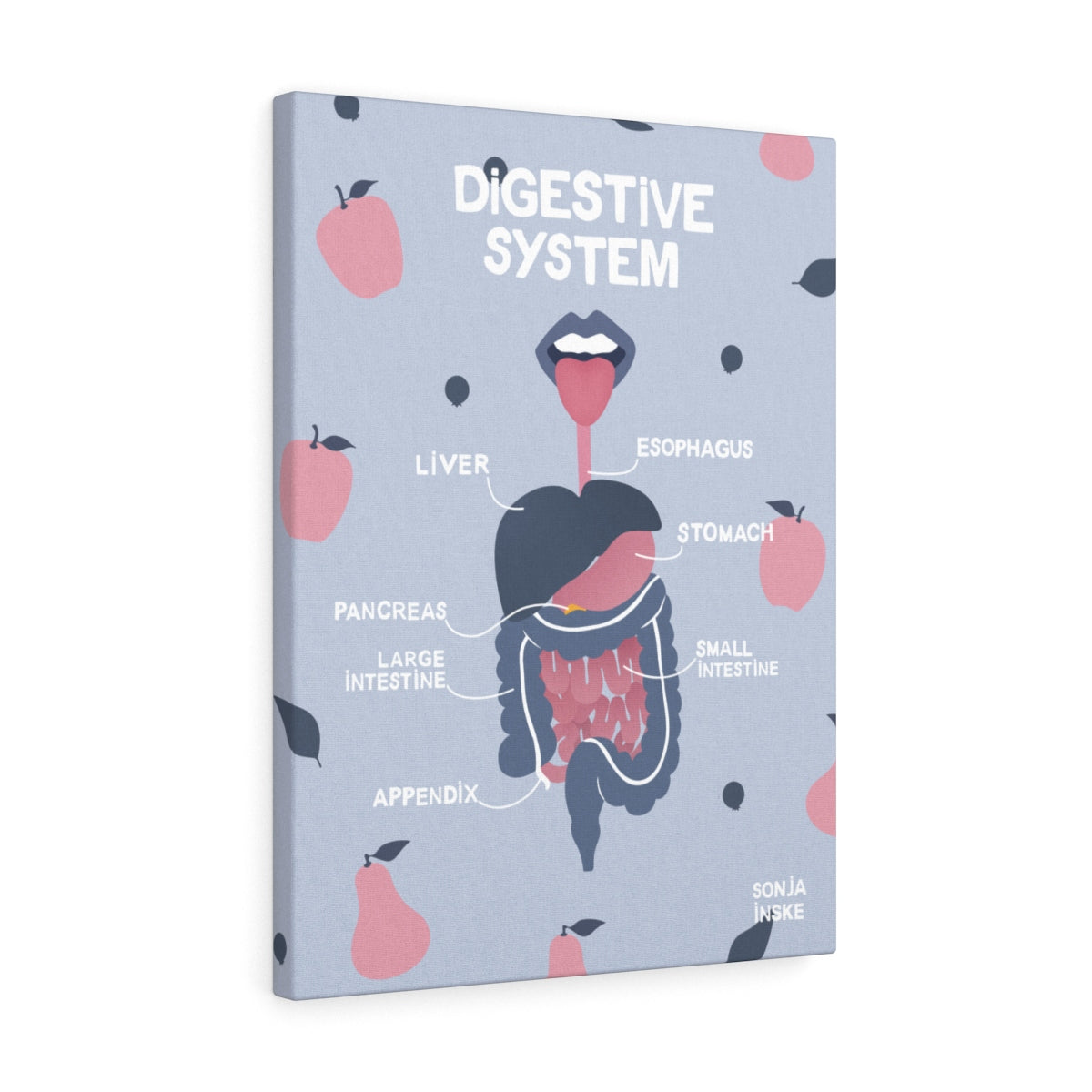
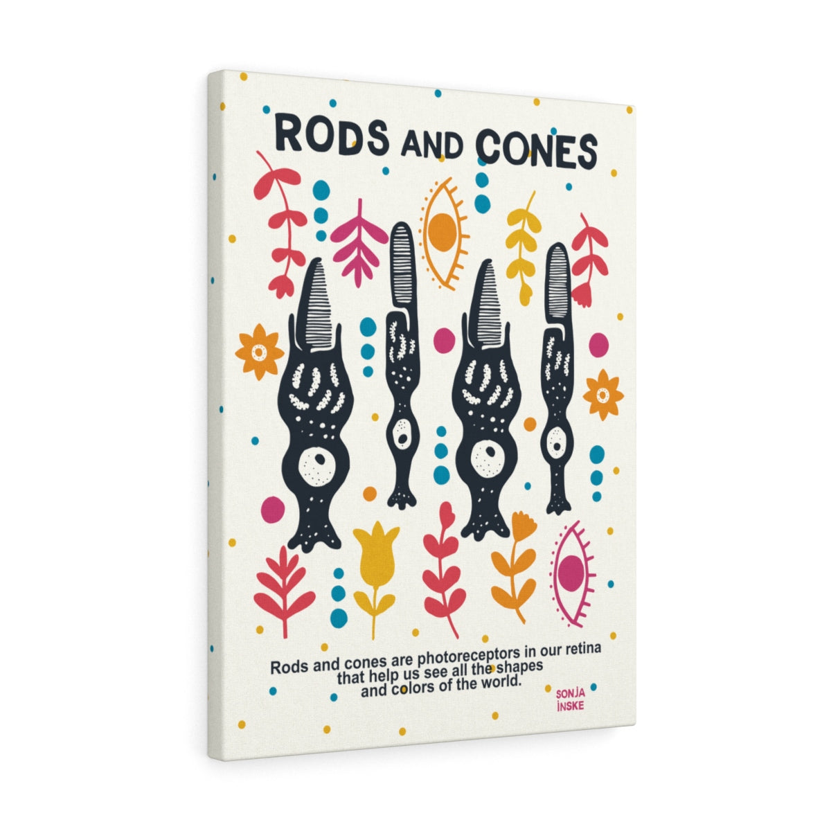
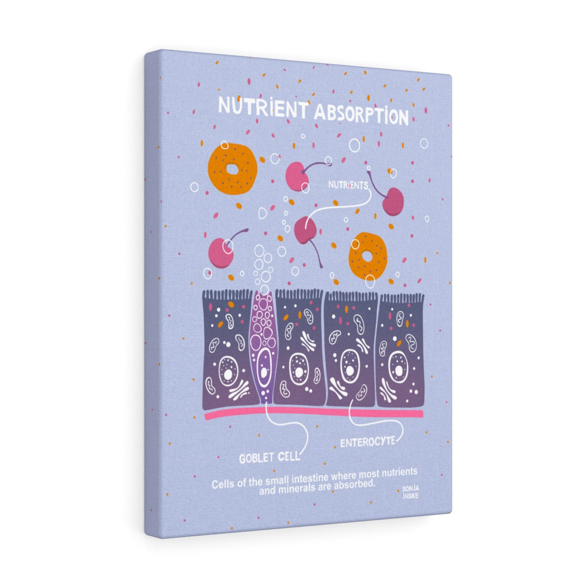
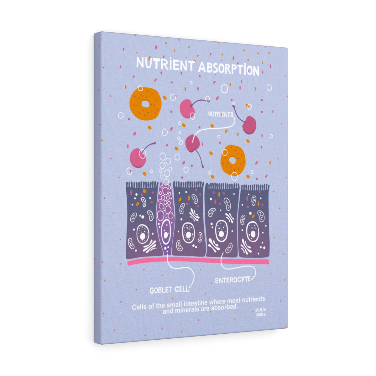
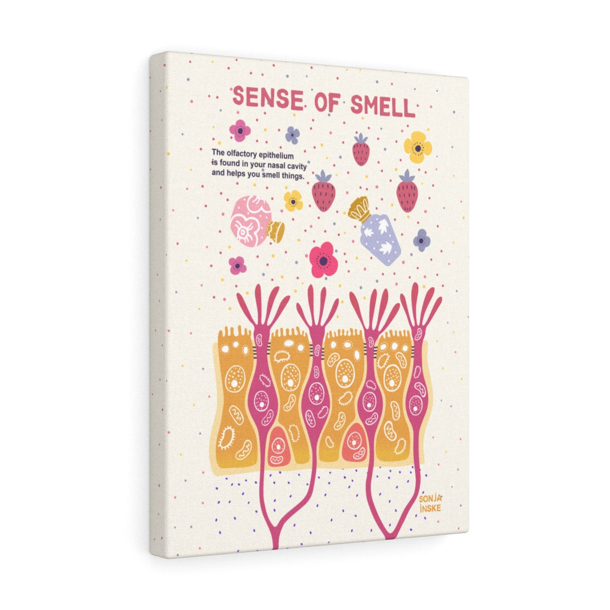
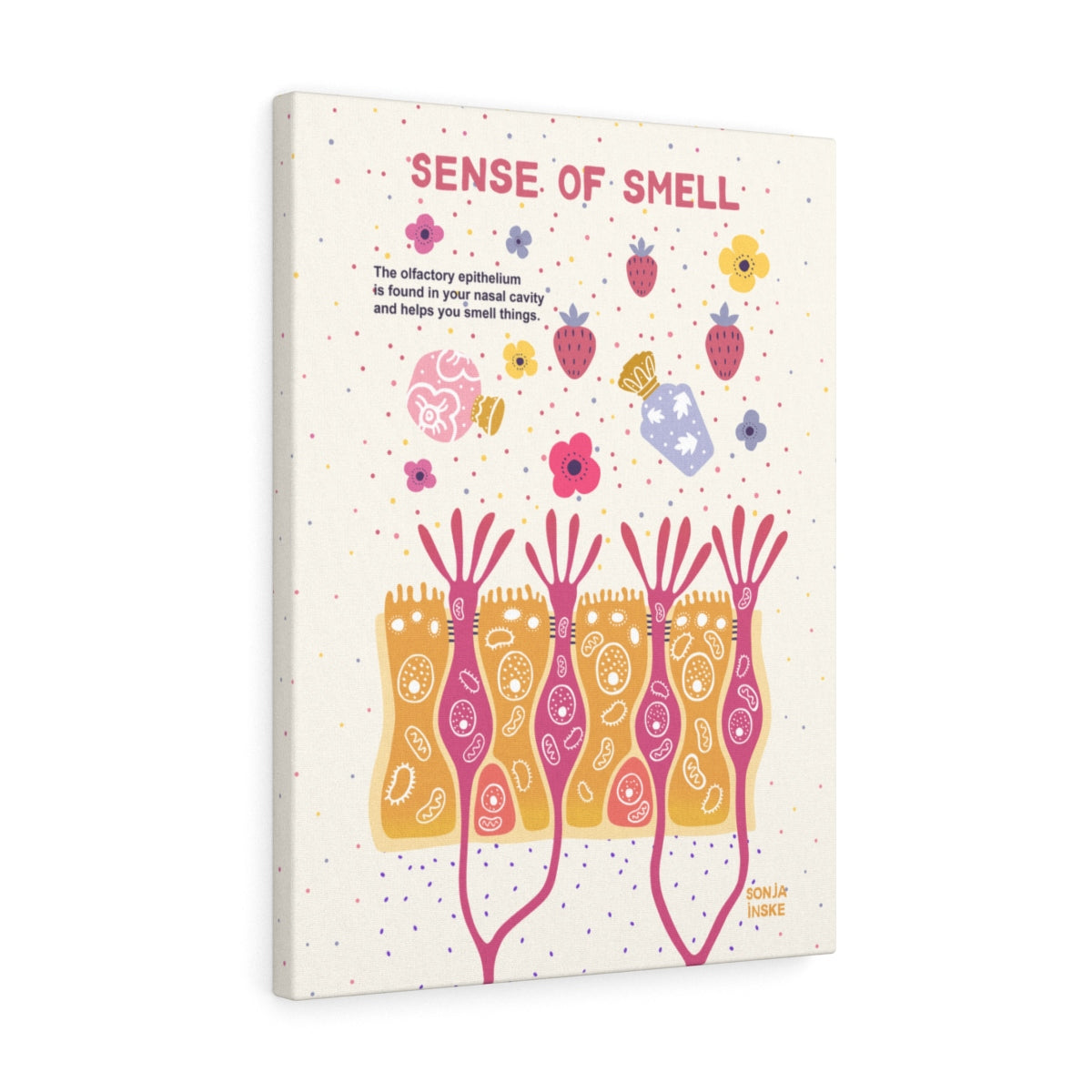
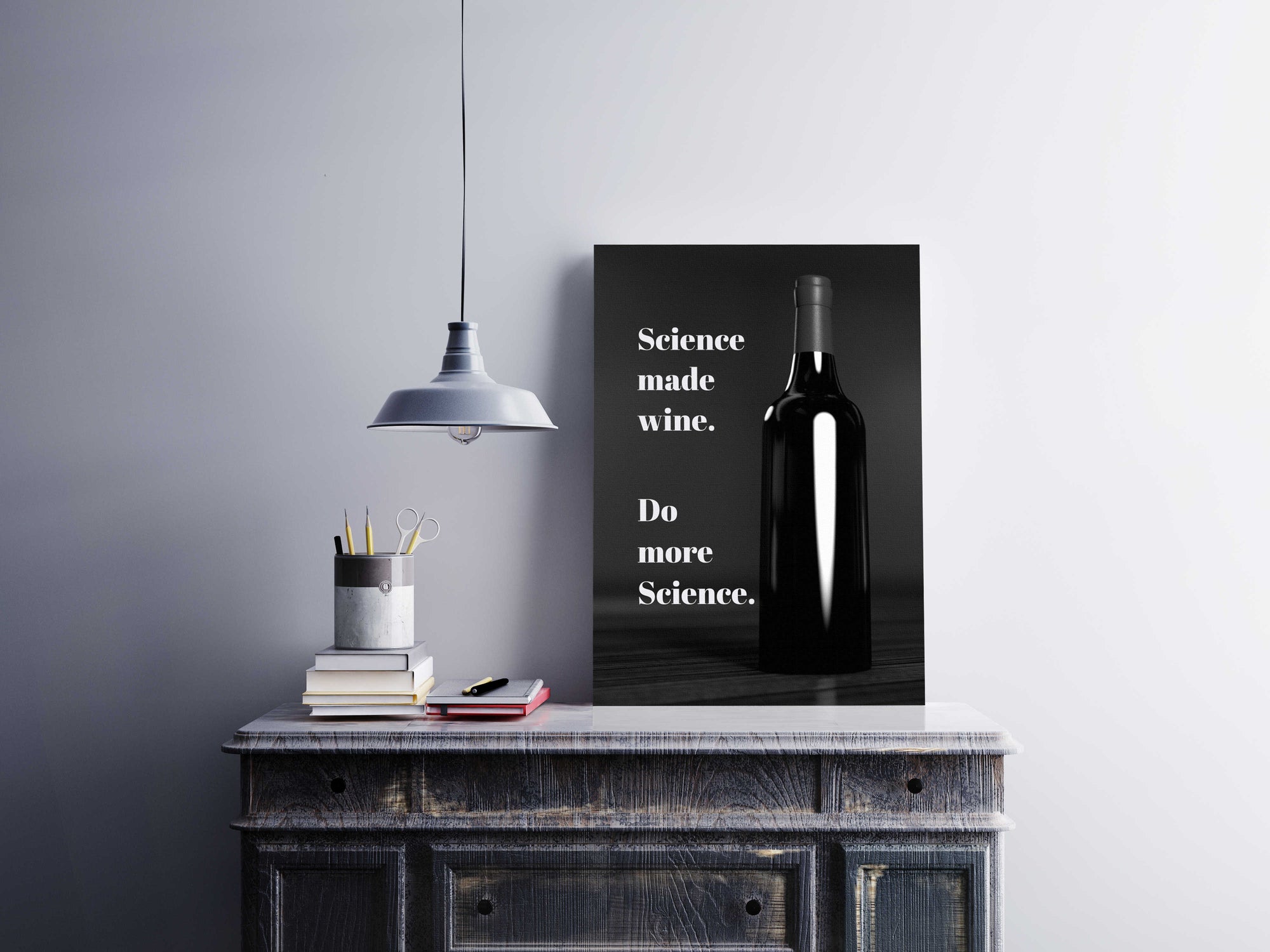
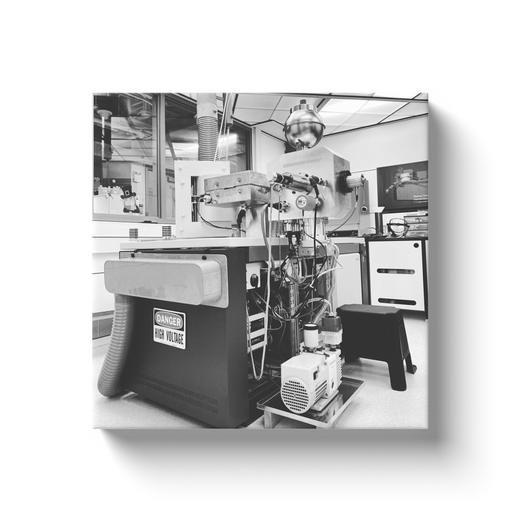
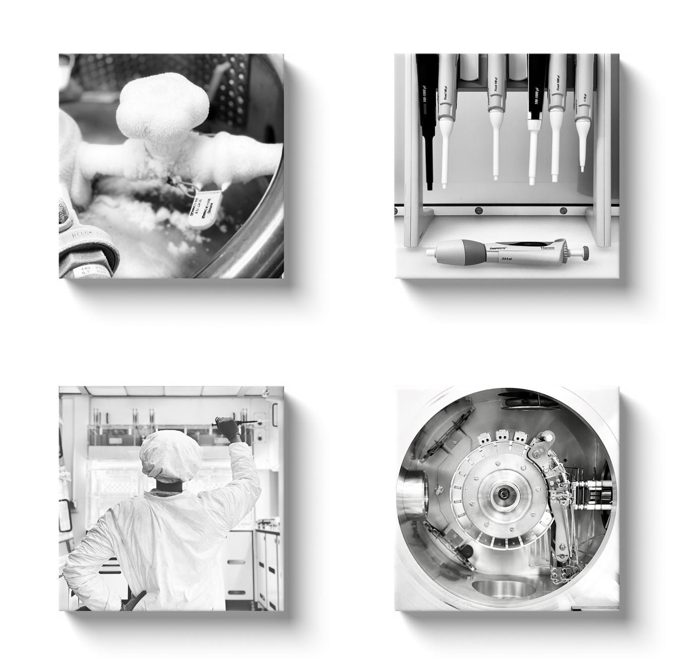
The biological world has a wealth of information and beauty that can be uncovered by a passionate scientist with the right tools.
This micrograph was captured by cardiovascular scientist Lindsey Avery Fitzsimons (@LAF_in_the_lab on Instagram and Twitter). Lindsey's research focuses on understanding congenital heart disease, the most common birth defect which affects about 1% of all children.
Create a welcoming waiting room, a colorful office or elevate the mood in your laboratory with science art that is both beautiful and informative. This biological science art is a perfect fit for a cardiologist office, family doctor or hospital exam room.
Image caption reads:
Left ventricle wall of an embryonic mouse heart at 63x magnification by Leica confocal microscopy
Blue: cell nuclei; Magenta: endothelial/endocardial cells; Green: tyrosine kinase/EGFR receptor
Captured by Lindsey Avery Fitzsimons, @LAF_in_the_lab - details at GeniusLabGear.com/LAF
The science behind the art:
(3D/Z-stack at 63Xoil/Zoom Factor 1.00) Confocal microscopy (Leica Microsystems) of a transverse (axial) frozen tissue section of the embryonic mouse heart at embryonic timepoint E12.5 (12.5 days post-fertilization); cross section has been collected at the atrioventricular level, and magnifies the left ventricular wall of the developing heart; blue/DAPI = cell nuclei; magenta/PECAM-1(CD-31)= endothelial/endocardial cells; green/ErbB4 = tyrosine kinase/EGFR receptor ErbB4 (Her4)
Product Details:
Made in the USA. Printed on museum-quality textured canvas with no glare or sheen. Frames are made from recycled plastic making them solid, durable, lightweight and environmentally friendly.
Pre-installed saw-tooth hangers | Scratch-resistant UV coating to prevent fading | Corner bumpers for level hanging and to protect your wall | Individually printed with attention to detail | Arrives in 1-3 weeks in the USA, 2-5 weeks internationally